Scientific Program

Conference Series Ltd invites all the participants across the globe to attend 2nd Global Pediatric Ophthalmology Congress Milan, Italy.

Day :

  • Retina & Retinal Disorders|Corneal Diseases
Speaker

Chair

Ronni M Lieberman

New York City Health and Hospitals Corporation, USA

Session Introduction

Ronni M Lieberman

New York City Health and Hospitals Corporation

Title: Long-term follow-up of intravitreal bevacizumab for the treatment of pediatric retinal and choroidal diseases

Time : 11:55-12:20

Speaker
Biography:

Ronni M Lieberman has received her Medical training at SUNY Downstate in New York, where she also completed her Ophthalmology Residency. She then went on to complete a 2 year Surgical Retina Fellowship at Mount Sinai Medical Center in New York. She is the Director of Medical Retina Services for the Queens Hospital Network, an HHC affiliate. She has published extensively, participated in clinical trials and is involved in the training of ophthalmology residents in the HHC and at Mount Sinai Medical Center.

Abstract:

The purpose is to describe the long-term outcomes of intravitreal bevacizumab (IVB) for the treatment of pediatric retinal and choroidal diseases. We performed a retrospective analysis of all pediatric patients receiving IVB at our HHC facilities for retinal and choroidal disease. We analyzed the data for visual acuity, stabilization of disease process and central macular thickness for 1 year. Safety data (adverse events) were analyzed. All eyes receiving IVB for the following were included in this analysis: Sickle cell retinopathy, Coats disease, familial exudative vitreoretinopathy (FEVR), SRNM (secondary to trauma, myopia) and CME secondary to uveitis of various etiologies. IVB was used both as a primary treatment and in combination with other options. Patients receiving IVB for ROP were analyzed separately. Patients receiving IVB for pediatric retinal and choroidal disease tolerated the treatment well. Most patients experienced significant improvement or stabilization of vision but long term safety concerns still remains.

Cristina Nitulescu

National Institute for Mother and Child Health, Romania

Title: Retinopathy of prematurity in Romania

Time : 12:20-12:45

Biography:

Cristina Nitulescu has been involved in the Romanian National Retinopathy of Prematurity (ROP) Program since 2012, and from the beginning she realized that the premature babies’ destiny can be changed with a better care and a good screening program. She organized the first National ROP Symposium in 2015 for neonatologists, nurses and ophthalmologists, supported by the International Agency for Prevention of Blindness (IAPB) and the Romanian Society of Ophthalmology, to improve the children’s care and the results of ROP screening and treatment. She coordinates a new project, supported by IAPB, to mentor four ROP Excellence Regional Centers across the country, to provide support for a better collaboration at regional level and for the effective implementation of the ROP guidelines. It is mandatory that the health providers to be aware about ROP prevention, the mainstay of health care through the world.

Abstract:

Statement of the Problem: Retinopathy of prematurity (ROP) is the leading cause of childhood blindness in the world, but ROP detected and treated early is one of the most cost-effective preventions in all of medicine.
Purpose: Analysis of challenges and gaps in ROP National Program in Romania.
Methodology & Theoretical Orientation: In Romania the treatment of threshold ROP is based on the international classifying criteria for ROP (International Classification of Retinopathy of Prematurity) and on the results of the Early Treatment for Retinopathy of Prematurity Study – ETROP. There are few ROP cases presented with different treatment approaches for “threshold stage”- laser therapy, intra-ocular injection with Bevacizumab (Avastin), vitreoretinal surgery, alone or combined the evolution after treatment, as well as the reasons for unfavorable result in some cases.
Findings: The disease completely regressed after treatment for all the babies screened and treated in time. All the babies who progressed to retinal detachment were referred too late for screening. The standard of neonatal care is critical with respect to the risk of ROP.
Conclusion & Significance: The most important step in treating ROP is a good screening program. Laser therapy is the “gold” standard treatment for threshold ROP. Intra-ocular injection with Avastin is a therapeutic option for aggressive cases and for sick infants. We need a pediatric vitreoretinal surgery unit in Romania. In Romania, there is need more ophthalmologists specialized for ROP screening. The lack of screeners is opening doors to telemedicine, to ensure that every child at risk receives effective screening and treatment. The results of the screening program depend on a large measure on a good collaboration with neonatologists, pediatricians and parents.

Biography:

Ashutosh Patel have completed his Medical degree (M.B.B.S.) in February 2013 at the Manipal University, Karnataka India. He had joined S.S. Institute of medical sciences & research centre, Davangere, India as junior resident in dept. of Ophthalmology in June 2015 & currently he working in the dept. of ophthalmology. He had presented a paper titled as “iris claw lens-an alternative treatment modality for monocular aphakia” at national conference in February 2017, Jaipur, India. He had also presented two posters titled as “A rare presentation of ocular tuberculosis- a case report” and “retinopathy changes in an anaemic patients with diabetes mellitus” at national conference. He had submitted his paper titled as “incidence & associated symptoms of convergence insufficiency among general population” to a index journal titled as “Ophthalmology and Allied sciences (OAS)”. His paper has been selected for publication In OAS VOL.3 NO.2 JUL-DEC 2017.

Abstract:

Statement of the Problem: Retinopathy of prematurity (ROP) is a potentially blinding eye disorder of childhood that primarily affects premature infants weighing 1250 grams or less, born before 31 weeks of gestation. ROP is vasoproliferative eye disorder characterized by abnormal vascular development of retina. Various other risk factors also contribute. The purpose of this study is to describe the incidence of ROP in various neonate intensive care units (NICU) and obtain information about risk factors associated with ROP and treatment modalities.
Methodology & Theoretical Orientation: Premature infant born at 36 weeks of gestation or younger or 2000 grams or smaller, were screened for ROP. 1350 babies were screened between October 2012 and March 2016. Screening was done with RETCAM and indirect ophthalmoscope using 28D volk lens under topical anesthesia and those requiring treatment were given laser treatment. All the babies were followed up till 40 weeks of corrected gestation or until retina matured and further follow up continued for those with ROP until resolved. 
Findings: In 1350 premature neonates, ROP was detected in 248 infants. Most of the babies were in the range of 26-34 weeks of gestation age at birth. Total 74 infants had pre threshold ROP, out of which 49 infants had Type 1 or high risk pre threshold ROP and were given laser treatment. Remaining 25 infants had Type 2 or low risk pre threshold ROP and were followed weekly.
Conclusion & Significance: ROP is one of the important causes of ocular morbidities and childhood blindness. The magnitude of ROP in our study is 18.37%. Response to laser therapy is encouraging.
Recommendation: Gestational age and low birth weight were the most significant risk factors for development of ROP, thus screening those with more than 1.5 kg and gestational age up to 36 weeks need to be considered.
 

Speaker
Biography:

Cosimo Mazzotta has done his MD from School of Medicine, University of Siena, Italy in the year 1997. He did a Specialist Diploma in Ophthalmology in 2001 and has received his PhD in Ocular Pathology in 2006. He introduced the Corneal Cross Linking in Italy in 2004 being awarded by the Italian Society of Ophthalmology. In 2009, he became the Assistant Professor of Corneal Surgery at Optometry School of University of Siena, Italy. Currently, he is an Assistant Professor of Corneal Pathology and Surgery at Post-graduate Ophthalmology School of Siena University and Director of the Siena International Crosslinking Center. His areas of interest include corneal cross-linking, keratoconus, corneal transplants, corneal dystrophies, cataract and refractive surgery, and in vivo confocal microscopy.

Abstract:

Purpose: To assess keratoconus (KC) progression in pediatric patients with allergies who tested positive to surface matrix metalloproteinase 9 (MMP-9) point-of-care tests versus negative patients.
Methods: Prospective comparative study including 100 stage I-II keratoconic patients, mean age 15. All patients underwent an anamnestic questionnaire for concomitant allergic diseases and were screened with the MMP-9 point-of-care test. Patients were divided into 2 groups: KC patients with allergies (KC AL) and KC patients without allergies (KC NAL). Severity of allergy was established by papillary sub-tarsal response grade (PSR) and keratoconus progression assessed by corneal tomography, and corrected distance visual acuity (CDVA) measurement in a 12-month follow-up.
Results: The KC AL group included 52 patients and the KC NAL group 48. In the KC AL group 42/52 of patients (81%) were positive to MMP-9 point-of-care test vs. 2 positive patients in the KC NAL group (4%). The KC AL group data showed a statistically significant decrease of average CDVA, from 0.155±1.1 to 0.301±1.2 log MAR (p<0.005) at 12 months; K max value increased significantly, from 50.2 D±2.7 to 55.2 D±1.9 on average. The KC NAL group revealed a slight keratoconus progression without statistically significant changes. Person’s correlation test showed a high correlation between K max worsening and severity of PRG in the KC AL group.
Conclusions: The study demonstrated a statistically significant progression of KC in pediatric patients with concomitant allergies, positive to MMP-9 point-of-care test vs. negative. A high correlation between severity of allergy and keratoconus progression was documented.

Speaker
Biography:

Elias F Jarade, MD. is the director of the Corneal, External Disease, and Refractive Surgery Services at Beirut Eye Specialist Hospital. Dr. Elias is a graduate of Harvard Medical School with 2 certificates of fellowship in cornea and refractive surgery from the Eye Center and Eye Foundation for Research, and The Massachusetts Eye and Ear Infirmary, Harvard Medical School. Dr. Elias is heavily involved in the practice and research of cornea, cataract, refractive surgery for the past 15 years with main interest in keratoconus. Has to his record more than 50 peer reviewed scientific papers and chapters. Also, presenter and invited faculty in the field of cornea and refractive surgery at international meetings. Board member for the “Journal of Refractive Surgery” “International Journal of Ophthalmology”, “International Advisory Board for the Saudi Journal of Ophthalmology”, Founder-general secretary of the Emirates Cornea and Refractive Surgery (ECRS) club, Guest editor for the “Journal of Ophthalmology

Abstract:

Aim: To evaluate the safety and visual outcome of intracorneal ring segment (ICRS) implantation followed by crosslinking in pediatric keratoconus patients.
Method: This retrospective study included pediatric patients (≤14 years) with keratoconus and poor corrected distance visual acuity (CDVA) that underwent ICRS implantation and crosslinking (CXL). ICRS were inserted under topical anesthesia after creating a corneal tunnel with an IntraLase femtosecond laser. Crosslinking was performed after one month subsequently. Records were reviewed and data were collected preoperatively, at 6 months, at 1 year, 2 years and 4 years postoperatively. Additionally, an 8-year follow-up of a 9-year old patient with ICRS implantation is reported, but is not included into the statistical analysis, since CXL was performed only 7 years later. For this patient ICRS were inserted manually under general anesthesia.
Results: 12 patients (17 eyes; 10 males, 2 females) aged 9-14 years (mean age 12.3 years) received ICRS implantation followed by CXL. Follow-up times ranged from 6 months to 8 years after surgery. At the 6-month follow-up, all eyes were evaluated, at the 1-year, the 2-year and the 4-year follow-up 11, 10 and 7 eyes were evaluated, respectively. At the 6-month follow-up, mean CDVA in comparison to preoperative levels improved significantly (p=0.001) from 0.300.19logMAR to 0.120.1logMAR, mean uncorrected distance visual acuity (UDVA) also improved significantly from 0.900.50logMAR to 0.430.31logMAR. A significant decrease in both keratometry readings and spherical equivalence (from -4.0 D to -1.56 D) was also noted after ICRS insertion. At the 1-year, the 2-year and the 4-year follow-up refractive values remained relatively stable in comparison to the 6-month follow-up, except for a minor but significant improvement in cylinder and, at 4 years, in UDVA. The patient with the 8-year follow-up also showed visual improvement and a stable cornea. All patients tolerated the surgery well and no intraoperative or postoperative complications were reported, except for one ring segment that had to be removed after two years due to vascularization and corneal thinning.
Conclusion: ICRS implantation is a safe and effective procedure for visual rehabilitation in children with keratoconus and poor CDVA.

  • Pediatric Ophthalmology & Research | Pediatric Optometry
Speaker

Chair

Bruce H Koffler

Koffler Vision Group, USA

Session Introduction

Danielle M Ledoux

Boston Children’s Hospital - Harvard University, USA

Title: Surgical esotropia in Down syndrome patients

Time : 10:30-11:10

Speaker
Biography:

Danielle M Ledoux is a Pediatric Ophthalmologist, specializing in management of pediatric patients with a diagnosis of Down syndrome. She has been working at the Boston Children’s Hospital - Harvard University since 2007. In 2015, she completed her specialized in Pediatric Eye Care, a practice of pediatric ophthalmology and optometry in Massachusetts, USA. She has participated in significant numbers of studies both prospective and retrospective with special interest in those that pertain to the population of people with a diagnosis of Down syndrome. She also serves as a Consultant for the National Down Syndrome Society, an American non-profit organization dedicated to support of people and families as well as education of the public about Down syndrome.

Abstract:

Purpose: There is an increased incidence of acquired strabismus in the Down syndrome population, more commonly resulting in esotropia than other deviations. We performed a retrospective chart review of the Down syndrome population with esotropia who had strabismus surgery to better understand the types of esotropia seen and the surgical outcomes.
Methods: We performed a seven year (2007–2014) retrospective chart review of all patients with a diagnosis of Down syndrome who had surgery for strabismus at a single, tertiary care center. Data including indications for surgery, preoperative and postoperative sensorimotor exam, presence of anomalous head posture, and type of surgery were recorded. Postoperative exam was at least one month after surgery. Surgical success was defined using three criteria: 1) orthotropia to an esotropia of less than, or equal to 8 prism diopters in primary position, 2) improvement of anomalous head posture, and 3) no further indication for surgery.
Results: 16 patients were identified who had Down syndrome and were surgically treated for esotropia. Of these, one was excluded due to follow-up of only two weeks. Preoperative findings of the remaining 15 patients included 8 (53%) with an A-pattern, 1 (7%) with a V-pattern, 1 (7%) with a head tilt dependent esotropia, 2 (13%) who had no assessment of pattern, and 3 (20%) with no pattern to the esotropia. Anomalous head postures were appreciated in 5 (33%) patients preoperatively, resolved in 4 patients postoperatively, and persisted in 1. In addition, 2 other patients had anomalous head postures only appreciated postoperatively. Of the 8 patients with an A-pattern esotropia, 4 were treated with only a bilateral medial rectus recession, 3 (75%) of who required a second surgery. Ultimately, 7 of the 8 patients achieved successful surgical outcomes. Overall surgical success in this study was 87% (13/15). Two patients were deemed surgical failures due to persistence of anomalous head posture and esotropia.
Conclusions: Surgical success was achieved in 87% of patients with Down syndrome and esotropia. We identified an increased incidence of A-pattern strabismus in the Down syndrome population, a finding not previously noted. All the three patients who required reoperation had A-patterns that were not addressed at the time of surgery. Ultimately, 88% of patients with A-pattern strabismus achieved successful surgical outcomes. Recognition of patterns preoperatively may help guide the surgical plan.

Speaker
Biography:

Abstract:

Myopia is an epidemic affecting an increasing portion of the worldwide population. In Asian countries, studies now show 80-90% of young teenagers have myopia, and in the USA the number of affected children has risen from 25 to 40% in the last 15 years. Ophthalmologists are now getting more involved in trying to slow the progression in a child by intervening early with a number of treatments that have been found to be effective. These included the use of atropine drops, bifocal glasses and contacts, increasing outdoor activity, and the use of orthokeratology molding lenses. The author will discuss the current understanding of why these lenses work and review the current literature. Most studies using this technique show a 35-50% reduction in axial length over a 2-5 year period.

Speaker
Biography:

Anna Laura Giacomin has completed her Medical degree in 1986 at the Padua University. She has joined as the Ophthalmological Specialist at the Padua University in 1991. In 1996, she joined in the Ophthalmology Unit of the Camposampiero PD Civil Hospital, responsible for the Ophthalmoplastic Service. She has obtained Homeopathy clinic course and in 2011, Master 2° level in Ophthalmoplastic Surgery from Naples Federico II University. From 2005, she is a full Member of the European Society of Ophthalmic Plastic and Reconstructive Surgery.

Abstract:
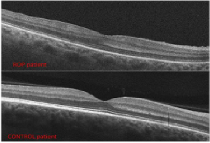
Introduction: Retinopathy of prematurity (ROP) can lead to blindness due to retinal detachment, but also premature infants with a normal apparent retina can develop subnormal visual acuity. Foveal depression and inner retinal layers develop mainly before the 40 week of gestational age (GA), while outer retinal layers develop mainly post birth. The purpose of our study was to correlate inner and outer foveal structural alterations with visual function and to correlate these alterations with GA, body weight (BW), and stage of ROP in preterm infants with ROP regressed spontaneously or after laser treatment.
Methods: Thirty eyes of 15 premature children aged 4 to 9 years with previous ROP underwent a complete ophthalmological and orthoptic evaluation, spectral domain OCT and angio-OCT (Canon OCT-HS100). The parameters evaluated with SDOCT in the fovea were: Total retinal thickness, inner retinal layers thickness, Henle’s layer thickness, outer nuclear layer thickness, external limiting membrane integrity, ellipsoid zone and outer segment tips zone integrity. Foveal avascular zone and foveal intra-retinal vascular plexus were evaluated with ANGIO-OCT. Other parameters evaluated were: Best corrected visual acuity (BCVA), refraction after cycloplegia. 10 eyes of 5 normal term children served as controls.
Results: Total central foveal thickness, inner retinal layer thickness, Henle’s layer thickness are significantly higher compared to term children. Outer nuclear layer thickness is lower compared to term children. Impaired visual function is correlated to impaired outer retinal layers structure. These changes are correlated to GA, BW, and stage of ROP.
Conclusions: A correct macular development at birth period is crucial for normal visual function. An impaired foveal cell migration and development can lead to a subnormal visual function in children with history of ROP. Further studies are necessary to understand the causes of foveal structure changes in preterms and the influence of macular edema of prematurity.

Lelio Sabetti

University of L’Aquila, Italy

Title: Revital Vision treatment in patients affected by nystagmus

Time : 16:10-16:35

Biography:

Lelio Sabetti is an Ophthalmologist who is in charge of the Pediatric Ophthalmology and Strabismus Unit at the Department of Biotechnological and Applied Clinical Sciences of the University of L’Aquila, Italy. Most of his efforts have concentrated on strabismus in children and adults, and treatment of amblyopia. He is also a Fellow of the Italian Strabismus Association (AIA) and of the European Strabismological Association (ESA).

Abstract:

Purpose: To evaluate the efficacy of the RevitalVision training in enhancing Best Corrected Visual Acuity (BCVA) and Contrast Sensitivity (CS) in a group of patients affected by nystagmus.
Materials & Methods: 12 patients, ranging in age from 10 to 45 years (median age 25.9), 9 males and 3 females with nystagmus and low vision: 5 cases with congenital nystagmus, 3 with albinism, 3 with congenital cataract (treated surgically) and 1 case with multiple sclerosis were studied. Each patient underwent a comprehensive ophthalmic examination, including measurement of visual acuity (BCVA) using LogMar ETDRS charts, contrast sensitivity using the FACT TEST. Moreover we carried out a reading speed test, an examination of the Extra Ocular Movement (EOM), a Cover Test at near and distance (CT), Stereopsis (arc/sec) and a biomicroscopy of the posterior segment. Each patient followed a rehabilitation protocol including the administration of two questionnaires (GH-12 and SF-36) before and after treatment in order to measure self-esteem, role limitations due to physical functioning, social functioning and mental health. Neurovisual rehabilitation training sessions, both individual and customized, lasting about 35 minutes each with either eyes opened in order to train the cerebral cortex by means of contrast stimulus with decreasing size (Gabor Patch) were done. The treatment program consisted of 50 sessions to be administered to the patient on alternate days (at least three times a week) for a total of about 100 days.
Conclusion & Significance: At the end of the treatment, all patients reported subjective improvement in their vision. The functional training using RevitalVision, therefore, appears to be a promising therapy for the functional visual enhancement also in patients with a high visual impairment.

Angela Malik

Sidra Medical and Research Center, Qatar

Title: Pediatric contact lens fittings: little eyes big challenges

Time : 16:35-17:00

Biography:

Angela Malik is an Optometrist who graduated in 1991 from the University of Waterloo, Canada. Her expertise lies in the field of Pediatric Contact Lenses and Low Vision. She has years of clinical and teaching experience, both in private practice and in hospital settings. She has set up the pediatric contact lens optometry service in two leading hospitals in the Middle East. She currently practices at Sidra Medical and Research Center, Doha.

Abstract:

Contact lenses have a variety of applications in the management of corneal pathology, amblyopia and refractive error for the pediatric population. A vast range of contact lenses are available but choosing the correct lens for complex pediatric fittings poses significant challenges. The techniques for fitting these complex cases will be discussed as well as methodologies for the clinician to recognize the applications where lenses would be a more beneficial treatment option. Visual improvement may be achieved with contact lenses, but it is not the only role that such devices play.

Darakshanda Khurram

Moorfields Eye Hospital Dubai, UAE

Title: Clinical outcomes of surgical management of subluxated lenses in children

Time : 17:00-17:25

Biography:

Darakshanda Khurram has interest in Pediatric Ophthalmology. Her area of expertise covers the clinical and surgical management of congenital cataracts and congenital glaucoma including augmented filtration procedures. She is an active researcher, publisher and presenter in her specialist field. She undertook a Postgraduate Fellowship with the Royal College of Surgeons, Glasgow, UK. She completed her Fellowship Training in Pediatric Ophthalmology and Strabismus at Great Ormond Street Hospital and Moorfields Eye Hospital, in London, UK.

Abstract:

Ectopia lentis is the displacement of natural lens of the eye. It can occur after trauma or could be a sign of serious systemic disease. In children, developmental or hereditary systemic conditions like Marfan syndrome, homocystinuria, Ehlers- Danlos syndrome, most commonly and hyperglycemia, sulphite oxidase deficiency, simple primary ectopia lentis and congenital aniridia syndrome less commonly among others, cause lens subluxation. When lens is displaced anteriorly it results in narrowing of the anterior chamber angle and pupillary block leading to either acute or chronic angle closure glaucoma. Posterior dislocation of the lens can lead to vitreous traction on the retina causing retinal detachment or with leaking lens proteins into the vitreous cavity can cause chronic vitritis and chorioretinal inflammation. Partial subluxation with more than two third zonular supports can be managed with appropriate spectacle or contact lens correction. Visually significant lens subluxation causes irregular astigmatism and lenticular myopia leading to significant anisometropic amblyopia. Surgical management of ectopia lentis involves number of challenges and options. With the refinement of surgical techniques and adjunctive capsular devices and intraocular lenses, the clinical outcomes for children under going surgical management for ectopia lentis have improved greatly. Lensectomy within the bag with limbal approach is considered safe and effective in children. Intra-scleral fixation of IOL haptics or the newer designs of iris supported IOLs have been reported with good results. The purpose of our study is to report a series of children who underwent surgery for visually significant subluxated lenses. We measured visual acuity, refractive error and recorded complications in children after insertion of Artisan iris-claw implants. In conclusion, iris-claw lenses are safe and effective method of treating aphakia after in the bag lensectomy, in children with ectopia lentis.

Gosevska Dastevska Emilija

University Eye Clinic, Macedonia

Title: Convergence insufficiency
Biography:

Abstract:

Introduction: Convergence insufficiency (CI) is a common binocular disorder in which the eyes do not work at near easily. CI is the leading cause of astenopia, transient blurred vision, diplopia, headaches. Difficulty sustaining near-visual function and abnormal fatigue.
Purpose: Detection and diagnosis of convergence insufficiency in the school children from 6-15 years old.
Material & Methods: 72 children from 6 – 15 years old, that have complained of blurred vision and/or headages, loss of comprehension after short periods of reading or performing close activities were examined at the University Eye Clinic in Skopje. The ocular and orthoptic examinations were done and it was found that 26 of them have convergence of insufficiency. Were it was necessasry, we gave them spectacle correction. In all diagnosted patient with CI we gave the orthoptic exercises.
Results: After the treatment of the convergence insufficiency by orthoptic procedures, between the great numbers of patients above symptoms were reduced or disappeared.
Conclusion: Convergence insufficiency disorder frequently goes undetected in school age children. Standard ophthalmic and orthopic examination will enable detection of this binocular disorder. Convergence insufficiency can be successfully managed and it will allow these patients more comfortable and better quality of life.

Biography:

Abstract:

Purpose: The purpose of this study was to compare the outcomes of medial rectus (MR) muscle pulley fixation and augmented recession in children with convergence excess esotropia and variable-angle infantile esotropia.
Methods: This was a prospective randomized interventional study in which children with convergence excess esotropia or variable-angle infantile esotropia were randomly allocated to either augmented MR muscle recession (augmented group) or MR muscle pulley posterior fixation (pulley group). In convergence excess, the MR recession was based on the average of distance and near angles of deviation with distance correction in the augmented group, and on the distance angle of deviation in the pulley group. In variable-angle infantile esotropia, the MR recession was based on the average of the largest and smallest angles in the augmented group and on the smallest angle in the pulley group. Pre- and postoperative ductions, versions, pattern strabismus, smallest and largest angles of deviation, and angle disparity were analyzed.
Results: Surgery was performed on 60 patients: 30 underwent bilateral augmented MR recession, and 30 underwent bilateral MR recession with pulley fixation. The success rate was statistically significantly higher (P=0.037) in the pulley group (70%) than in the augmented group (40%). The postoperative smallest and largest angles and the angle disparity were statistically significantly lower in the pulley group than the augmented group (P<0.01).

  • Refractive Errors and Management | Pediatric Neuro-Ophthalmology | Ophthalmology Surgery
Speaker

Chair

Mikhail G Kataev,

Fyodorov Eye Microsurgery Complex, Russia

Session Introduction

Pedro Mattar

King Khaled Eye Specialist Hospital, Saudi Arabia

Title: Surgical management of ocular motor nerve palsies

Time : 11:30-11:55

Speaker
Biography:

Pedro Mattar is a Consultant at the Pediatric Ophthalmology and Strabismus Division at King Khaled Eye Specialist Hospital in Saudi Arabia. He has studied Pediatric Ophthalmology at University of Colorado, Denver, USA, Ophthalmology from AVAO Venezuela, and Medicine from Universidad Central de Venezuela. He is a member of American Association of Pediatric Ophthalmology and Strabismus, American Society of Cataract and Refractive Surgery, Venezuelan Society Ophthalmology, Pan American Association of Ophthalmology, Latin-American Pediatric Ophthalmology Society and Pan American Society of Retinopathy of Prematurity.

Abstract:

Ocular motor nerve palsies includes third, fourth and sixth nerve palsies. Congenital or acquire, can affect the adult and pediatric population; moderately or severely disturbs the position and movements of the eyes. Compensatory head posture, amblyopia and diplopia are among the clinical manifestation. High percentage is associated with systemic and neurological disease. After an intensive workup and a concise diagnosis a pediatric and strabismus specialist has the challenge to offer surgical treatment for alignment and diplopia free field in these patients. We present different surgical approaches in the management of ocular motor nerve palsies.

Biography:

Serap Azizoglu has completed her PhD from La Trobe University and currently works at Deakin University, School of Medicine, Deakin Optometry as Associate Lecturer in Optometric Clinical Skills.

Abstract:

Access to general health and eye care is related to an individual’s socioeconomic status (SES). We aimed to examine the prevalence of oculo-visual disorders in children in Istanbul Turkey, drawn from schools at SES extremes but geographically nearby. Three school-based vision screenings (presenting distance visual acuity, cover test, eye assessment history, color vision, gross stereopsis and non-cycloplegic autorefraction) were conducted on 81% of a potential 1014 primary-school children aged 4-10 years from a government (low SES) and two nearby private (high SES) schools in central Istanbul. Of the 823 children, mean age was 6.7±2.2 years; approximately 10% of all children were referred for a full eye examination (8.2% and 16.3% of private/government schools, respectively). Nearly 65% and 22% in the government/private schools respectively had never been previously examined. Of all children, 94.5% and 86.6% were able to read/identify with each eye the 6/9.5 or the 6/6 line of letters/shapes respectively, 7.9% presented wearing spectacles, 3.8% had impaired color vision, 1.5% had grossly impaired stereo-vision, 1.5% exhibited strabismus, 1.8% were suspected to have amblyopia and 0.5% reduced acuity of likely organic origin. Of the 804 without the latter three conditions, 6.0% were myopic ≤-0.50 DS, 0.6% hyperopic ≥2.00 DS, 7.7% astigmatic ≥1.00 DC and 6.2% anisometropic ≥1.00 DS. The results highlight the different pattern of visual problems associated with lifestyle differences in two populations raised in the same urban locale but drawn from different socioeconomic background and the need for general vision screenings prior to school entry .

Mikhail G Kataev

Fyodorov Eye Microsurgery Complex, Russia

Title: Oculoplasty for saving vision in children

Time : 12:20-12:45

Speaker
Biography:

Mikhail G Kataev is involved in Oculoplastic Surgery since 1978 after graduating from the 2nd Moscow Medical Institute. The beginning of ophthalmic surgery practice was set in Moscow Helmholtz Research Institute of Ophthalmology. In 2015, he became the Head of the Oculoplastic department at the Fyodorov Eye Microsurgery Complex, Russia. The range of surgical interest includes congenital, traumatic, senile, postmorbid pathology and aesthetic surgery as well. He has also supervised PhD theses related to ultrasonic surgery, carbon implants, surgical expanders, oriental eyelids and primary reconstruction in severe eyelids trauma. He is a member of the European Society of Plastic and Reconstructive Surgery since 1994.

Abstract:

Background: Normal morphology and function of ocular adnexa and orbit are critical in maintaining perfect vision. Early age is a most vulnerable period for developing vision function in children. Impairment of vision due to external ocular problems can be caused by epicorneal opacity or by the lack of seeing because of irritation, blepharospasm, epiphora, ptosis or strabismus.
Materials & Method: The whole cohort of more than 7000 pediatric oculoplastic patients in our data base contains about 4000 individuals susceptible to vision impairs. Oculoplastic surgery was indicated in cryptophthalmos, congenital symblepharon, and epicorneal opacity after burn, SJS, toxic epidermal necrolysis (TEN) causes direct blurring of the vision field. Potential risk of low vision (amblyopia) was an indication for surgery in strabismus, ptosis, and secondary blepharospasm, caused by conjunctival metaplasia, trichiasis, distichiasis, and entropion. The third indication was lagophthalmos due to eyelids skin deficit, ectropion or coloboma. All possible types of surgery were used to solve adnexal problems. Percent distribution in this specific group showed as follows: ptosis surgery 57%, strabismus surgery 17%, reconstruction of eyelids and fornices 21%, trichiasis 3% and orbital surgery 3%.
Results: The effect of treatment was evaluated by eyelids shape, proper mucous lining of the fornices, sufficient opening and closure of the eye fissure. In 98% of patients, anticipated result was achieved. Multistep surgery was often needed as a normal way of treatment. No serious complications were observed.
Conclusion: The effect of oculoplastic procedures corresponds differently with regaining of vision function. The correlation depends on the type of pathology. Ptosis and common strabismus repair leads to good outcome. Elimination of trichiasis and dysplasia of the conjunctiva facilitates vision and improvement in function. Meanwhile, restoration of the fornices in TEN patients suffering from dry type of lesion may stay poor. Nevertheless high rate oculoplastic surgery is a powerful tool for restoration of ocular adnexa and providing proper background enabling function repair.

Biography:

Ayad Shafiq began his training as a Paediatrician and changed specialisations to train as an Ophthalmologist with a special interest in Children s eye conditions, and inherited eye problems. Special interests include; Childrens eye problems from birth, Adult cataract surgery (approximately 450 operations per year for cataract) and Adult squint or strabismus.

Abstract:

Statement of the Problem: Systemic absorption of bevacizumab is a major concern in premature babies whose developing organs and brain may be adversely affected by suppression of vascular growth factors.
Rationale: We assessed the contralateral effect on the untreated eye by documenting serial images of the retinal vessels. Could even a micro-dose of bevacizumab have a demonstrable effect on the untreated eye by systemic vascular absorption? Most babies with APROP have simultaneous bilateral treatment. The asymmetry in this case allowed a short period of careful observation which could provide important qualitative evidence of systemic drug effect.
Type of Study: This was an observational study of a single case.
Methodology: Both eyes were photographed prior injection, at day 5 and at day 12. Images were compared to assess dilatation and tortuosity in both treated and untreated eyes.
Results: Both eyes responded to injection of one eye. A smaller but lesser reduction was noted in the untreated eye in vascular tortuosity and dilatation of posterior pole vessels.
Conclusion: Even a micro-dose of 0.16 mg bevacizumab injected in one eye, has a qualitative effect on retinopathy of prematurity in the untreated eye. Systemically absorbed bevacizumab appears to be absorbed adequately to have an end organ effect. The 'standard' dose currently used in premature babies is 0.625 mg, four times greater dose than that used in this baby. We treat APROP in all cases with a micro-dose of bevacizumab. This is effective in a case series of bilateral primary treatment. Systemic absorption has been pharmacologically proven for a larger standard dose of bevacizumab. This study adds evidence to the suggestion that even a micro-dose has systemic end organ effects. In view of the unknown potential systemic effects, research should be directed at identifying the smallest dose which is effective at preventing blindness in APROP.

Speaker
Biography:

Elias F Jarade, MD. is the director of the Corneal, External Disease, and Refractive Surgery Services at Beirut Eye Specialist Hospital. Dr. Elias is a graduate of Harvard Medical School with 2 certificates of fellowship in cornea and refractive surgery from the Eye Center and Eye Foundation for Research, and The Massachusetts Eye and Ear Infirmary, Harvard Medical School. Dr. Elias is heavily involved in the practice and research of cornea, cataract, refractive surgery for the past 15 years with main interest in keratoconus. Has to his record more than 50 peer reviewed scientific papers and chapters. Also, presenter and invited faculty in the field of cornea and refractive surgery at international meetings. Board member for the “Journal of Refractive Surgery” “International Journal of Ophthalmology”, “International Advisory Board for the Saudi Journal of Ophthalmology”, Founder-general secretary of the Emirates Cornea and Refractive Surgery (ECRS) club, Guest editor for the “Journal of Ophthalmology”.

Abstract:

Purpose: To report the incidence, clinical presentation, risk factors, and treatment outcome of pediatric keratoconus in a tertiary referral eye hospital in Beirut, Lebanon.
Methods: In this retrospective study, the authors evaluated all patients with keratoconus 14 years or younger newly diagnosed at the Beirut Eye Specialist Hospital, Beirut, Lebanon, between January 2010 and December 2014. The incidence of pediatric keratoconus among all pediatric patients and among patients with keratoconus of all ages was assessed. Patients with pediatric keratoconus were evaluated for keratoconus stage, initial presentation, uncorrected distance visual acuity, corrected distance visual acuity (CDVA), corneal topography, and pachymetry. Patients were classified according to different treatment regimen groups and different follow-up visits were evaluated. 
Results: During 5 years, 16,808 patients were examined, of whom 2,972 were 14 years or younger. A total of 541 patients were diagnosed as having keratoconus; of those, 16 were 14 years or younger at the time of diagnosis. Hence, the incidence of keratoconus was 0.53% among pediatric patients and 3.78% among adult patients (>14 years). Initial presentation was during routine checkup (1 of 16) for allergic conjunctivitis (3 of 16), reduced vision (10 of 16), and corneal hydrops (mimicking keratitis) (2 of 16). Except for 2 patients lost to follow-up, all eyes received corneal cross-linking treatment and 16 eyes received additional intracorneal ring segment implantation.
Conclusions: The incidence of pediatric keratoconus indicates that increased awareness for keratoconus among children is needed, mainly in cases of family history of keratoconus, ocular allergy/pruritus, poor CDVA, corneal hydrops, and/or high astigmatism.

Biography:

Lelio Sabetti is an Ophthalmologist who is in charge of the Pediatric Ophthalmology and Strabismus Unit at the Department of Biotechnological and Applied Clinical Sciences of the University of L’Aquila, Italy. Most of his efforts have concentrated on strabismus in children and adults, and treatment of amblyopia. He is also a Fellow of the Italian Strabismus Association (AIA) and of the European Strabismological Association (ESA).

Abstract:

Aim: To investigate the effects of choline combined with Bangerter filter in the treatment of amblyopia and to evaluate increase in visual acuity. The evaluation was conducted in comparison to patients undergoing treatment with Bangerter filters alone.
Methods: A sample of 80 amblyopic subjects with initial average visual acuity of 0.63 with follow-up at 12 months was studied. All subjects were assigned to use a Bangerter filter placed on the corrective spectacle lens all waking hours and alternating the eye (1 day over the left eye, 1 day over the right eye). Choline was administered orally to 39 patients once daily, five days per week for the entire study period. Patients underwent ophthalmic examination of the anterior and posterior segment of the eye to assess the acuity vision in LogMar and refraction in cyclopegia, orthoptic examination with ocular motility study, cover/ uncover test with prism and fusion range.
Results: All patients demonstrated an increase in visual acuity. The patients with exodeviation associated with the administration of choline showed a 50% improvement of vision at 12 months and a 44.79% improvement was observed in patients with esodeviation. The subjects assigned to the Bangerter group experienced improvement in vision at 12 months, specifically a 28% improvement in patients with exodeviation and a 34% improvement in subjects with esodeviation was observed.
Conclusions: The filter is an effective amblyopia treatment; however these effects are markedly enhanced when coupled with the administration of choline. Findings suggest that the effects are particularly relevant in the more severe amblyopic cases.

Antonella Vecchies

University of Padua, Italy

Title: Management of scleral lenses in pediatric age

Time : 14:35-15:00

Biography:

Antonella Vecchies has done her Master’s degree in Vision Science from the University of Padua. She is an Optometrist and has done her specialization in contact lenses for the irregular cornea and in pediatric population. She has also worked at the Children's Hospital Burlo Garofolo (Trieste-Italy).

Abstract:

Scleral lenses are large diameter rigid gas permeable lenses that range from 14 mm to over 20 mm in diameter. Scleral lenses completely cover the cornea and extend onto the sclera and are supported by the conjunctival tissue. Pediatric patients with keratoconus, pellucid marginal degeneration, post-penetrating keratoplasty, corneal scarring or ocular surface disorders are most appreciative of the benefits of scleral lenses. In cases of ocular surface disease or persistent epithelial defects, scleral lens wear can facilitate healing and corneal health. The goals of scleral lenses fitting in pediatric age are visual rehabilitation for refractive errors, protection the ocular surface and reducing symptoms in severe ocular surface disease. The benefits of scleral lenses are a great stability and centration on ocular surface without any touch in the cornea. To obtain corneal clearance the recommended overall lens diameter is at least 2 mm larger than the corneal diameter. It is necessary that the sagittal depth of the initial lens selected be greater than the sagittal height of the cornea. The elevation of an individual cornea may be measured using topographical elevation data, anterior segment OCT imaging. The OCT permits to evaluate with a great precision central corneal clearance, limbal clearance and haptic zone.

Biography:

He is an Orthoptist of the European Reference Network Center for Low Vision. In 2005, he got general certificate of Education State Secondary School Lyceum specializing in scientific and technological studies “G. B. Quadri” of Vicenza from Italy. In 2008, he completed his degree in Orthoptics and Opththalmologic Assistence from School of Medicine, University of Padua, Italy and from 2009 – 2010 Study Course and Master in Optometry from Institute B. Zaccagnini – Bologna, Italy. In 2017 he Study Course and Training Program entitled: “Visual Training in Educational, Dyslexia and Sport Vision” from Scientific Institute Intervision – Milan, Italy.

Abstract:

Learning disabilities are a common problem in the pediatric population and are generally associated to memory dysfunction or mathematical calculations. Efficient reading is accomplished through complex and interrelated processes, one of which is vision. Determining the relationships between vision and learning involves more than evaluating eye health and visual acuity it is a multidisciplinary approach (ophthalmologist, orthoptist/optometrist, posturologist and logopedist) in which all appropriate areas of function are assessed and managed. Current research indicates that some people with reading difficulties, such as difficulties related to dyslexia, have co-existing visual and language processing deficits. Unresolved visual deficits can impair the ability to respond fully to educational instruction. To identify learning related vision problems we need to fully evaluate the three interrelated areas of visual function: 1) Visual pathway integrity including eye health, visual acuity and refractive status; 2) Visual efficiency including accommodation (focusing), binocular vision (eye teaming) and eye movements; and 3) Visual information processing including identification and discrimination, spatial awareness, and integration with other senses. Efficient reading requires accurate eye movements and continuous brain integration of the information obtained from each eye. The full diagnosis and management of many oculomotor anomalies (ocular dominance, fixation, vergence amplitude, saccade and smooth pursuit) helped by automatic computerized tests are necessary to increase learning ability simply by identifying the one missing micro-skill. Visual training is a method attempting to correct or improve presumed ocular disorders, visual processing, and perceptual disorders. Vision therapy can be broadly divided into two categories. In the first category, classic orthoptic techniques are used to correct accommodative (focusing) and convergence dysfunctions as well as heterophorias (latent misaligned eyes) and refractive errors (need for glasses) that might be responsible for asthenopic symptoms (eye fatigue and discomfort often aggravated by close work). In the second category, often referred to as behavioral vision therapy, eye movement and hand-eye coordination training techniques are used to improve visual processing skills, learning efficiency, and visual-motor integration. Behavioral vision therapy is based on the premise that differences in children’s visual perceptual motor abilities exist and that these perceptual motor abilities influence cognitive and adaptive skills such as reading, writing, and motor activities used in activities of daily living. Behavioral vision therapy has been recommended to improve visual skills and processing in the belief that this will improve learning disabilities, including speech and language disorders, and nonverbal learning disorders. In conclusion, the management plan of patient with dyslexia and learning disabilities include treatment, guidance and appropriate referral. The expected outcome is an improvement in visual function with the alleviation of associated signs and symptoms. Vision therapy does not directly treat learning disabilities or dyslexia but improve visual efficiency and visual processing, becoming a part of a multidisciplinary approach thereby allowing the person to be more responsive to educational instruction.

Oscar Orjeda

Peru

Title: Glaucoma children in Peru

Time : 15:25-15:50

Biography:

Oscar Orjeda is a Medical Ophthalmologist, graduated from the Universidad Mayor de San Marcos and is a Ophthalmology Specialist at the Cayetano Heredia University. He has done his specialization in Pediatric Ophthalmology in Pittsburgh, and in Glaucoma from Argentina. He is a Doctor at the Children's Hospital, Peru. He is the Medical Director and General Manager of the Optima Vision Clinic, Peru. He is also a Principal Investigator of Glaucoma in Children at Parexel International, USA.

Abstract:

In the present study we evaluated the phenotype of the Peruvian inhabitant and compared it in relation to childhood glaucoma. The study was carried out from May 1997 to May 2015. We evaluated 94 patients that were equivalent to 169 eyes where 52% were primary childhood glaucoma and 48% were secondary childhood glaucoma. We compared the cases of Vander Helm (1965), Sampaolesi (1991) and ours both in gender and bilaterality, concluding that in relation to gender, unlike the two authors mentioned that give a ratio of 70% for men and 30% for women, we found a ratio of 55% for men and 45% for women; with respect to present symptoms, 100% of cases present photophobia, with 5% of cases of rhinorrhea. The type of camerular angle that the patient presents according to Sampaolesi, which finds a Type I angle in 70% of cases and Type II in 30%, we found 60% of cases for an angle Type I and 40% of cases for a Type II angle. In the use of anti-metabolites, we found that 5 fluorouracil produces small and highly vascularized blisters unlike the blisters produced by mitomycin which are flat avascular blisters. In relation to valve implants, we found that adult Ahmed implants could be used without any problem in children's eyes. We conclude that ocular pathology is closely related to the ethnology of each region.

Speaker
Biography:

Elias F Jarade, MD. is the director of the Corneal, External Disease, and Refractive Surgery Services at Beirut Eye Specialist Hospital. Dr. Elias is a graduate of Harvard Medical School with 2 certificates of fellowship in cornea and refractive surgery from the Eye Center and Eye Foundation for Research, and The Massachusetts Eye and Ear Infirmary, Harvard Medical School. Dr. Elias is heavily involved in the practice and research of cornea, cataract, refractive surgery for the past 15 years with main interest in keratoconus. Has to his record more than 50 peer reviewed scientific papers and chapters. Also, presenter and invited faculty in the field of cornea and refractive surgery at international meetings. Board member for the “Journal of Refractive Surgery” “International Journal of Ophthalmology”, “International Advisory Board for the Saudi Journal of Ophthalmology”, Founder-general secretary of the Emirates Cornea and Refractive Surgery (ECRS) club, Guest editor for the “Journal of Ophthalmology”.

Abstract:

Purpose: To evaluate the safety and visual outcome of intracorneal ring segment (ICRS) implantation followed by crosslinking in pediatric keratoconus patients.
Design: Retrospective interventional case series
Methods: This retrospective study included pediatric patients (≤14 years) with keratoconus and poor corrected distance visual acuity (CDVA) that underwent ICRS implantation and crosslinking (CXL). ICRS were inserted under topical anesthesia after creating a corneal tunnel with an Intralase femtosecond laser. Crosslinking was performed 1 month subsequently. Records were reviewed and data collected preoperatively, at 6 months, at 1 year, 2 years and 4 years postoperatively. Additionally, an 8-year follow-up of a 9-year old patient with ICRS implantation is reported, but is not included into the statistical analysis, since CXL was performed only 7 years later. For this patient ICRS were inserted manually under general anesthesia.
Results: 12 patients (17 eyes; 10 males, 2 females) aged 9-14 years (mean age 12.3 years) received ICRS implantation followed by CXL. Follow-up times ranged from 6 months to 8 years after surgery. At the 6-month follow-up all eyes were evaluated, at the 1-year, the 2-year and the 4-year follow-up 11, 10 and 7 eyes were evaluated, respectively. At the 6-month follow-up, mean CDVA in comparison to preoperative levels improved significantly (p=0.001) from 0.300.19logMAR to 0.120.1logMAR, mean uncorrected distance visual acuity (UDVA) also improved significantly from 0.900.50logMAR to 0.430.31logMAR. A significant decrease in both keratometry readings and spherical equivalence (from -4.0D to -1.56D) was also noted after ICRS insertion. At the 1-year, the 2-year and the 4-year follow-up refractive values remained relatively stable in comparison to the 6-month follow-up, except for a minor but significant improvement in cylinder and, at 4 years, in UDVA. The patient with the 8-year follow-up also showed visual improvement and a stable cornea. All patients tolerated the surgery well and no intraoperative or postoperative complications were reported, except for one ring segment that had to be removed after two years due to vascularization and corneal thinning.
Conclusion: ICRS implantation is a safe and effective procedure for visual rehabilitation in children with keratoconus and poor CDVA.

Goshevska Dashtevska Emilija

University Eye Clinic, Macedonia

Title: Treatment of anisometropic amblyopia
Biography:

Abstract:

Introduction: Anisometropic amblyopia is the second most common cause of amblyopia. Anisometropic amblyopia can occur when there is a difference in refractive error between the two eyes.
Purpose: The purpose of this study is to evaluate factors predicting success in the treatment of anisometropic amblyopia.
Materials & Method: The records of 18 children from 3-8 years old, treated successfully for anisometropic amblyopia were reviewed. Optimal refractive correction was provided. Age, initial visual acuity and stereoacuity of anisometropia were analyzed. The time course of improvement in visual acuity and the factors related to amblyopia resolution were assessed. Patching or atropine penalization was considered in additional to optical management if patients show no improvement with glasses alone after 3 months.
Results: 18 children with a mean age of 5.6 years were included. Mean time to amblyopia resolution was 5.8 months (range 2 to 15 months). Worse best corrected initial visual acuity was associated with longer time to resolution. Seven (38.9%) of the hyperopic patients achieved visual acuity of 20/20 in the investigated period. Better initial stereoacuity predicted good final stereoacuity. Treatment outcome was not related to age, but was related to better baseline visual acuity and lesser amounts of anisometropia.
Conclusion: Compliance with treatment has major effect on response to therapy. Treatment of anisometropic amblyopia with glasses alone can be a successful option. Patching or pharmacological penalization can be added if there is no improvement in visual acuity only with glasses after 3 months.