Koshits Ivan
Petercom-Network / Management Systems Consulting Grope Cl. Corp., Russia
Title: Theory-Physiological and biomechanical features of the interconnected functioning of the systems of accommodation, and aqueous production and outflow. Hypotheses and actuating mechanisms of growth of the eye’s optical axis in the metabolic theory of adaptive myopia and in the theory of retinal defocus
Biography
Biography: Koshits Ivan
Abstract
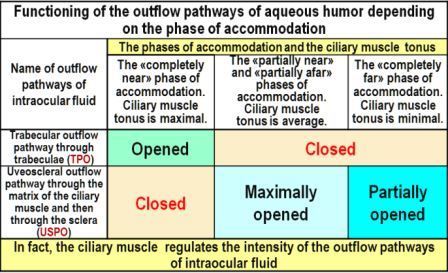
1. Physiological features of the interconnected functioning of the systems of accommodation, and aqueous humor production and outflow: The regulation systems for accommodation, production and outflow of aqueous humor have one common actuating unit – the ciliary muscle (CM), both in animals and humans. At the same time, the control signals from these systems to the CM can be directly opposite [4,6,8]. For example, to open the trabecular pathway of outflow (TPO), it is necessary to reduce the CM. But at the same time, it is necessary to relax the CM in order to see the approaching danger in the distance. So which command will be executed first? In the eye, there is an overriding priority for executing commands from the accommodation system, since the survival of the species as a whole depends on this. The signals of the control system of aqueous production are in the second place by priority, and the signals from the outflow control system are performed last. It is because that the task of maintaining metabolic processes in the eye is more important than the task of removing the spent aqueous humor [4,6,17]. Such physiological representations are key to understanding the features of the inte rrelated functioning of these three physiological systems of the eye. Most of the animals have only one aqueous outflow pathway – uveoscleral pathway of outflow (USPO), which then passes into the outflow through the sclera. Only in humans and in four species of highly evolved monkeys, during the course of evolution, an additional aqueous outflow pathway was formed through the trabeculae (TPO, trabeculae outflow pathway). This happened because of changes in the habitat, which required to develop the ability of a long visual work at a short distance, and at that moment the USPO is blocked (in the ciliary muscle, the interfiber spaces with the matrix are compressed at that moment). In Table 1 it is shown that TPO is open only when looking near, and USPO is closed at that time. In visual work at medium and long distances, on the contrary, only USPO is open, and TPO is closed. It should be noted that USPO is the main way by which the necessary ingredients are delivered to maintain normal metabolism and reproduction of collagen in the middle and back parts of the sclera. Also, along this basic pathway, prostaglandins are delivered to the sclera, which are normally produced by the intraocular epithelium. The sclera can regulate its permeability with the help of a large number of prostaglandin receptors located in it [1]. That is why the pharmacotherapy of glaucoma with prostaglandins is so effective. The eye does not control the level of IOP directly, since morphologists have not yet detected baroreceptors in it. The level of IOP in the eye is directly determined primarily by the level of rigidity of the sclera [2-4]. A large number of mechanoreceptors have been found in the sclera [1], which allow to control the reciprocal displacement of scleral plates during micro fluctuations of the eye volume. Conclusion: the eye does not control the level of IOP, but constantly monitors its volume with the help of mechanoreceptors, as well as the receptors of prostaglandins [4].

The outflow (slow filtration) of the aqueous humor occurs through the three main eye filters: juxtacanicular tissue, inter-fiber ciliary matrix and scleral matrix. The outflow efficiency is determined by the main functional characteristic of the sclera – its fluctuability (this is a new concept in ophthalmology). Fluctuation is the functional ability of the sclera to "push out" the waste intraocular fluid from the eye with the help of elastic fibers and fibroblasts located in the sclera. Concurrently the volume of the eye decreases. We have learned to reliably measure in vivo the level of fluctuation and rigidity of sclera, as well as the level of IOP in youth and even in elderly patients, using an ORA air analyzer by our own method [2-4].
2. The key links of adaptive myopia (AM) <br>
The development of the most pandemic myopia in the history of mankind shows that until recently there was no workable theory of acquired myopia in the world. The forecast of an increase in the number of myopes to 5 billion people by 2050 (half of the world's population, figure 1) [5] shows that if acquired myopia is a disease, it is transmitted "visually". But the clinical facts show another pattern. AM develops rapidly both in animals and in a healthy person under the age of 45 years. And this apparently is a normal adaptive reaction, which provides the possibility of prolonged strenuous visual work at a short distance. Adaptive myopia is a clear example of the implementation of the energy saving law in anatomical development of biological systems. The length of the optical axis of the eye should be such as to ensure the necessary long-term near visual work with minimal tonus of the ciliary muscle [4,6]. Since children of all animals and humans at an early age are weak hyperops, even the transition to emmetropia or initial myopia can significantly reduce the energy consumption of the eye if necessary to perform long-term near work. Highly developed monkeys, such as gorillas, orangutans and others, are even forced to post a sentry along the perimeter of the troop, because their way of life implies gathering food and looking at it closely.

