Day 1 :
Keynote Forum
Janette Atkinson
University College London, UK
Keynote: Cerebral visual impairment: dorsal stream vulnerability from amblyopia and autism to attention
Time : 10:05-10:35

Biography:
Janette Atkinson is an internationally renowned pioneer in research on normal and abnormal visual development in infants and young children, directing Visual Development Unit at Cambridge University, UCL London University and Oxford University. Together with collaborators in ophthalmology, visual neuroscience, optometry and pediatric neurology, she has broken new ground in devising novel visual tests and techniques to understand visual brain development and used these findings to assess and remediate visual impairment. She has over 150 peer-reviewed publications in leading international journals. She has been honored by election to fellowships of the Academy of Medical Sciences, Academia Europaea, and the British Academy and awarded Kurt Koffka Medal of Giessen University for research in visual science. In addition, she was awarded Davida Teller Award of Vision Sciences Society in 2016 for her outstanding contribution to vision research.
Abstract:
Advances in assessing sensory and cognitive vision in infants and young children have enabled us to establish milestones of visual development and provide means for identifying visual disorders at an early age. These include novel EEG and behavioural techniques and tests such as Cambridge Crowding cards, the first child-friendly preschool crowded acuity test for identifying amblyopia, and photo- and video refraction (VPR1), for rapid identification of refractive errors at any age. Using these new techniques, in our population RCT refractive screening programs of over 8000 infants, we demonstrated that refractive correction with glasses in infancy could successfully reduce the incidence of strabismus and amblyopia. However, many children who had been significantly hyperopic in infancy showed mild pre-school visuomotor and attentional deficits. Combined with similar deficits in neurodevelopmental disorders, this led us to devise measures of development of the cortical dorsal stream, underpinning motion sensitivity, visuomotor control of actions and eye movements, and the ventral stream, underpinning orientation, shape, and object/face perception. The ball in the grass test compares motion sensitivity (dorsal) with static form sensitivity (ventral). We found deficits in motion sensitivity in children with Williams syndrome, autism, very preterm birth, perinatal brain injury and hemiplegia. This dorsal stream vulnerability (DSV) has now been found in many disorders, both genetic and acquired, including amblyopia. Besides poor motion sensitivity, it includes deficits in visuomotor control and visual attention. From MRI, we find that children’s individual differences in motion sensitivity are correlated with specific areas of growth in parietal cortex, the integrity of the superior longitudinal fasciculus (major fibre tract connecting frontal with parietal areas) and visuomotor and mathematical ability. This suggests that motion sensitivity relates to higher-order decision-making and visual attention, and may provide useful early diagnostic indicators of visuo-cognitive deficits in amblyopia, cerebral visual impairment and genetic retinal dystrophies.
- Special session on “From East to West of Europe: Different approaches in pediatric ophthalmologyâ€
Location: London, UK

Chair
Andreea Ciubotaru
Infosan Eye Clinic, Romania
Session Introduction
Andreea Ciubotaru
Infosan Eye Clinic, Romania
Title: Reoperations in Duane retraction syndrome

Biography:
Ciubotaru Andreea completed her Medical degree at Carol Davila University of Medicine and Pharmacy, Romania in 1998 and; Ophthalmology Residency at Emergency Eye Hospital, Bucharest from 2000–2005. In 2011, she completed her PhD at Ludwig Maximilian University Munich, Germany, with the thesis entitled “The superior oblique posterior tenectomy as therapy for congenital Brown’s syndrome”. Since 2007, she is the Medical Director of Infosan Eye Clinic Bucharest; President of Romanian Society of Pediatric Ophthalmology and Strabismus and Member of Romanian Society of Cataract and Refractive Surgery and Board Member of Romanian Society of Ophthalmic Genetics. She contributes as author in many Romanian and international congresses and also books of ophthalmology. Her preferred topics are strabismus, amblyopia, congenital cataract, and ophthalmic genetics.
Abstract:
Introduction: Patients with strabismus who have undergone previous eye muscle surgery often present a challenge to strabismus surgeons, especially in restrictive strabismus cases like Duane’s retraction syndrome (DRS). Even has been a recognized clinical entity for nearly a century, DRS is sometime mistaken for essential infantile esotropia or abducens paralysis. <br>
Methods: We retrospectively evaluated 15 patients undergoing surgery for esotropia 4 to 22 years ago in other eye clinics. All patients were referred to us for the misalignment in primary position and abnormal head position. After the previous operation, nine patients developed a consecutive exotropia (group 1) and six of them were still esotropic in primary position (group 2). Angle
of strabismus in primary position, head posture, abduction and adduction of the affected eye were measured before and after surgery in our clinic. Photos from childhood were reviewed and the diagnosis for all was DRS. <br>
Results: 12 patients have first heard about DRS. In group 1, the intraoperative findings showed a previous medial rectus recession (in the affected eye) in six cases and a contralateral medial rectus recession (in the normal eye) in three cases. The mean deviation in primary position decreased from 28 prism diopters (PD) exotropia preoperatively to eight PD exotropia postoperatively. In group 2, the intraoperative findings showed in the affected eye a previous medial rectus recession in three cases and lateral rectus resection in three cases. The mean deviation in primary position decreased after reoperation from 25 PD esotropia preoperatively to 10 PD esotropia postoperatively. All patients improved their abnormal head posture after reoperation. <br>
Conclusion: Although the clinical findings and the surgical approach in Duane’s retraction syndrome are very clear, in medical practice from Romania we meet even today misinterpreted forms of DRS requiring reoperation.
Iosub Ionela
Infosan Eye Clinic, Romania
Title: Inferior oblique recession is enough for unilateral long-standing congenital superior oblique palsy?

Biography:
Abstract:
Introduction: Congenital superior oblique palsy (CSOP) is sometime overlooked by the general ophthalmologist due to the absence of manifest vertical deviation (VD) in primary position (PP), even if it has been a recognized clinical entity for nearly a century.
Methods: We retrospectively evaluated 46 adults undergoing surgery for unilateral CSOP in our clinic (2006-2017). The patients were divided in two groups: Group 1-32 patients with preoperative VD in PP less than 30 prism diopters (PD), in which we performed a graded inferior oblique recession (8 mm, 10 mm or maximal) and group 2-14 patients with preoperative VD in PP greater than 30 PD, in which we performed a maximal inferior oblique recession combined with recession of contralateral inferior rectus muscle. Horizontal and vertical deviations in five positions of gaze and head posture were assessed pre- and postoperatively.
Results: In group 1, VD in PP and in adduction was reduced from median 10 PD and 25 PD to 1 PD and 3PD respectively. In group 2, VD in PP and in adduction was reduced from median 36 PD and 34 PD to 8 PD and 8 PD respectively. All patients improved their abnormal head posture after reoperation. 78% of patients have first heard the diagnosis of CSOP in our clinic; even 92% of them had more than three eye examinations during life.
Conclusion: Although the clinical findings in CSOP are very clear, in medical practice from Romania we meet even today overlooked forms of CSOP, even with massive VD in PP.
Andreea Hopinca
Infosan Eye Clinic, Romania
Title: Superior oblique posterior tenectomy in congenital brown's syndrome

Biography:
Abstract:
First described 60 years ago, congenital Brown’s Syndrome ( a disorder of ocular motility manifesting most notably a restriction of active and passive elevation in adduction) is even up to now under debate regarding its etiopathogenesis and surgical methods. This paper reviews the various etiologic theories, from anomalies of the superior oblique tendon and/or the trochlear apparatus to recent hypothesis that congenital Brown’s syndrome might represent a congenital cranial dysinnervation syndrome, caused by a fourth nerve hypoplasia or aplasia with consecutive paradoxical innervation of the superior oblique muscle. Most used surgical treatments for Brown’s syndrome are described and evaluated, with accent on superior oblique posterior tenectomy, a surgical approach used also in Infosan Clinic.
- Retina & Retinal Disorders | Corneal Diseases
Location: London, UK

Chair
Ronni M Lieberman
New York City Health and Hospitals Corporation, USA
Session Introduction
Ronni M Lieberman
New York City Health and Hospitals Corporation, USA
Title: Evidence based use of intravitreal avastin and laser in ROP: Developing a new treatment algorithm

Biography:
Ronni M Lieberman completed her Medical degree at SUNY Downstate in New York, and then Ophthalmology Residency. She completed her two-year fellowship
in Medical and Surgical Retinal Disease at Mt Sinai Medical Center, in New York. She is the Director of Ophthalmology at Queen Hospital Center, both a New York City Health and Hospital Corporation and the Icahn School of Medicine, Mt Sinai Medical Center affiliate. Her main interests include Infectious Retinitis and ROP. She has published papers extensively on a number of topics in retina and vitreous.
Abstract:
The talk will discuss the etiology and staging of retinopathy of prematurity (ROP) and various treatment options. It will then focus on newer algorithms of combination therapies in the face of long term follow up. This will include our experience at the HHC/Mt Sinai using these techniques.
Boris I Gramatikov
Wilmer Eye Institute, Johns Hopkins University School of Medicine, USA
Title: A computer system integrating central fixation detection and optical coherence tomography for pediatric applications

Biography:
Boris I Gramatikov has his areas of expertise which includes “Electronics, optoelectronics, computers, computer modeling, signal/image processing, data analysis, electronic instrumentation design, bio-photonics, ophthalmic optics, and biomedical optics, all applied to the development of diagnostic methods and devices for ophthalmology and vision research”. His team has developed a series of pediatric vision screeners. Along with Dr. Cynthia Toth from Duke University, he is co-PI on a project for combining optical coherence tomography with retinal birefringence scanning, to enable OCT imaging in toddlers and young children. He has over 120 publications, 37 of which in high-impact peer-reviewed journals. He serves as a Reviewer and Editorial Board Member of a number of technical and medical journals.
Abstract:
Optical coherence tomography (OCT) enables volumetric rendering and the generation of fundus images that precisely and register OCT images to fundus features. Yet, there is little data about how a child’s retina develops. This limits
our knowledge of how diseases affect a child’s vision early in life and makes diagnosis of these diseases more difficult. The introduction of OCT to pediatric applications has been impeded by several factors, among them data acquisition and analysis speed, and difficulty in attaining stable fixation of the pediatric patient on a target over a period of time long enough to allow reliable analysis. The system described here integrates three major components: a computer-controlled video player that plays attention attracting movies and directs the subject’s fixation to a central point target; a retinal birefringence scanning subsystem for fast detection of central fixation by detecting the position of the fovea, and; an optical coherence tomography subsystem for acquiring 3D images from the retina (mainly in the foveal region). From a main menu, the operator selects a suitable video to be played by a small monitor in the visual field of the child under programmatic control. The software then starts the birefringence scanning system that scans the area around the fovea using polarized light, and in a double-pass configuration, analyzes the changes of polarization caused by the Henle fibers around the fovea. This allows fast detection of central fixation. The OCT system is instructed by the central software to acquire data if and only if central fixation is detected. This significantly reduces data redundancy and the time needed for analysis of the OCT data, while improving the overall reliability of the system. The idea is applicable to handheld devices.
Camelia-Margareta Bogdanici
University of Medicine and Pharmacy, Romania
Title: Amblyopia-A challenge for ophthalmologist and parents

Biography:
Camelia-Margareta Bogdanici completed her General Medicine at University of Medicine and Pharmacy Grigore T Popa, Romania in 1985. She did her Residency from 1990-1994, in the same hospital in which she is working now. She was an Assistant Professor at University of Medicine and Pharmacy Grigore T Popa, first in Histology department from 1987-1990; in Ophthalmology department from 1991-1999; Lecturer from 1999-2007 and; then an Associate Professor in the same university and hospital. Her lectures are for Romanian and English students in Ophthalmology. She completed Medical Degree (PhD) in 1999. The title of her thesis was “Clinical research for pressure equilibrium at normal, ocular hypertension and glaucoma”. From 2007-2012, she was Medical Director of Hospital Sf. Spiridon Iasi, Romania, and from 2012-2016, she was Deputy in Romanian Parliament, in Health and Family Commission. She is the member of Romanian Board Society, Romanian Society of Ophthalmopediatry and Strabismus, member of Romanian Society of Contact Lenses, member of European Academy of Orthokeratology. She contributes as author in many Romanian and International Congresses, and also of scientific publications and books of ophthalmology.
Abstract:
Amblyopia is an important condition that can affect up to 5% of the general population. Ophthalmologist has many difficulties both in amblyopia diagnosis and treatment. Difficulties of diagnosis can be produced by missing: of children cooperation in examination, of parent’s sincerity in history of the child (or even of treatment done before), of medical, a busy medical program or even missing of amblyopia experience. Difficulties of treatment can be: the moment of diagnosis, small compliance from children or parents or small experience of doctor. It is not possible to state that the impact of amblyopia treatment felt by the child is the same of the adults. The parents can only make a judgment on how they perceive the child is able to see whilst on treatment. Their judgment could be influenced by how important they judge the activity to be. Treatment compliance in amblyopia therapy is influenced by both the child and the parents. Whilst the child may object to the wearing of glasses or a patch on a personal level, a parent’s perspectives can influence the success of such treatment. This may incorporate their own experiences of patching/glasses-wear, or their understanding of the condition and the importance of treatment.
Parental understanding of the condition and belief in the prescribed treatment are key components for good treatment compliance. An argument against using treatment compliance as a measure of quality of life is that a child may consent to wearing the patch but their daily activities and social interactions may still be affected. Currently in Romania there is high interest in preventing amblyopia. For these ophthalmologist tries to inform population doing visual screening for children and by informing through media and press.
- Interdisciplinary Symposium “Physiology and biomechanics of the eyeâ€
Location: London, UK

Chair
Svetlova Olga
Russia
Session Introduction
Koshits Ivan
Petercom-Network / Management Systems Consulting Grope Cl. Corp., Russia
Title: Theory-Physiological and biomechanical features of the interconnected functioning of the systems of accommodation, and aqueous production and outflow. Hypotheses and actuating mechanisms of growth of the eye’s optical axis in the metabolic theory of adaptive myopia and in the theory of retinal defocus

Biography:
Abstract:
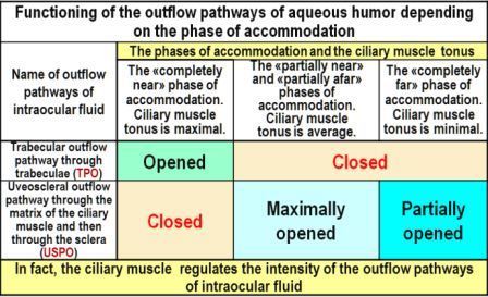
1. Physiological features of the interconnected functioning of the systems of accommodation, and aqueous humor production and outflow: The regulation systems for accommodation, production and outflow of aqueous humor have one common actuating unit – the ciliary muscle (CM), both in animals and humans. At the same time, the control signals from these systems to the CM can be directly opposite [4,6,8]. For example, to open the trabecular pathway of outflow (TPO), it is necessary to reduce the CM. But at the same time, it is necessary to relax the CM in order to see the approaching danger in the distance. So which command will be executed first? In the eye, there is an overriding priority for executing commands from the accommodation system, since the survival of the species as a whole depends on this. The signals of the control system of aqueous production are in the second place by priority, and the signals from the outflow control system are performed last. It is because that the task of maintaining metabolic processes in the eye is more important than the task of removing the spent aqueous humor [4,6,17]. Such physiological representations are key to understanding the features of the inte rrelated functioning of these three physiological systems of the eye. Most of the animals have only one aqueous outflow pathway – uveoscleral pathway of outflow (USPO), which then passes into the outflow through the sclera. Only in humans and in four species of highly evolved monkeys, during the course of evolution, an additional aqueous outflow pathway was formed through the trabeculae (TPO, trabeculae outflow pathway). This happened because of changes in the habitat, which required to develop the ability of a long visual work at a short distance, and at that moment the USPO is blocked (in the ciliary muscle, the interfiber spaces with the matrix are compressed at that moment). In Table 1 it is shown that TPO is open only when looking near, and USPO is closed at that time. In visual work at medium and long distances, on the contrary, only USPO is open, and TPO is closed. It should be noted that USPO is the main way by which the necessary ingredients are delivered to maintain normal metabolism and reproduction of collagen in the middle and back parts of the sclera. Also, along this basic pathway, prostaglandins are delivered to the sclera, which are normally produced by the intraocular epithelium. The sclera can regulate its permeability with the help of a large number of prostaglandin receptors located in it [1]. That is why the pharmacotherapy of glaucoma with prostaglandins is so effective. The eye does not control the level of IOP directly, since morphologists have not yet detected baroreceptors in it. The level of IOP in the eye is directly determined primarily by the level of rigidity of the sclera [2-4]. A large number of mechanoreceptors have been found in the sclera [1], which allow to control the reciprocal displacement of scleral plates during micro fluctuations of the eye volume. Conclusion: the eye does not control the level of IOP, but constantly monitors its volume with the help of mechanoreceptors, as well as the receptors of prostaglandins [4].
The outflow (slow filtration) of the aqueous humor occurs through the three main eye filters: juxtacanicular tissue, inter-fiber ciliary matrix and scleral matrix. The outflow efficiency is determined by the main functional characteristic of the sclera – its fluctuability (this is a new concept in ophthalmology). Fluctuation is the functional ability of the sclera to "push out" the waste intraocular fluid from the eye with the help of elastic fibers and fibroblasts located in the sclera. Concurrently the volume of the eye decreases. We have learned to reliably measure in vivo the level of fluctuation and rigidity of sclera, as well as the level of IOP in youth and even in elderly patients, using an ORA air analyzer by our own method [2-4].
2. The key links of adaptive myopia (AM) <br>
The development of the most pandemic myopia in the history of mankind shows that until recently there was no workable theory of acquired myopia in the world. The forecast of an increase in the number of myopes to 5 billion people by 2050 (half of the world's population, figure 1) [5] shows that if acquired myopia is a disease, it is transmitted "visually". But the clinical facts show another pattern. AM develops rapidly both in animals and in a healthy person under the age of 45 years. And this apparently is a normal adaptive reaction, which provides the possibility of prolonged strenuous visual work at a short distance. Adaptive myopia is a clear example of the implementation of the energy saving law in anatomical development of biological systems. The length of the optical axis of the eye should be such as to ensure the necessary long-term near visual work with minimal tonus of the ciliary muscle [4,6]. Since children of all animals and humans at an early age are weak hyperops, even the transition to emmetropia or initial myopia can significantly reduce the energy consumption of the eye if necessary to perform long-term near work. Highly developed monkeys, such as gorillas, orangutans and others, are even forced to post a sentry along the perimeter of the troop, because their way of life implies gathering food and looking at it closely.

Marina Guseva
City diagnostic medical center "Vodokanal of St. Petersburg", Russia
Title: The art of choosing rational optical correction using the eyeglasses and contact lenses of modern design in the light of the metabolic theory of adaptive myopia

Biography:
Marina G. Guseva: From 2003 to 2011 Chief doctor-ophthalmologist of 54 urban centers "Nevskaya Optics holding», with 2011 Chief ophthalmologist «Sfera» LTD, with 2012 ophthalmologist in Medical city center "Vodokanal of St. Petersburg". Have 9 international certificates for optometry. Author of 34 publications, co-author of two chapters in the book "Ophthalmocontactology" (St. Petersburg: Military-medical Academy, 2010). Co-author and developer of the modern way of rational optical correction and specialized training manual "Physiological principles of rational optical correction. Practical recommendations. Normal physiology.
Abstract:
Statement of the Problem: Fighting adaptive myopia (AM) will only be effective when truly working theory of myopia is developed and confirmed by clinical trials. The pandemic of myopia confirms the absence of a theory that can explain all known clinical facts and give physiologically approved practical recommendations. And as practice has shown, unfortunately, the incremental retinaldefocus theory (IRDT) is no exception. The hypothesis of this theory not always corresponds to the known clinical facts about the physiology of the eye taking into consideration interconnected operation of accommodation and aqueous humor production and outflow systems [1]. However, in 2001 Russian authors Ivan Koshits (biomechanics) and Olga Svetlova (ophthalmologist) proposed a metabolic theory of adaptive myopia (MTAM), which was based on a profound long-term interdisciplinary analysis of modern clinical facts in such fields like morphology, physiology, biomechanics and accommodative mechanisms of the eye [1]. MTAM not only explains known clinical facts, but also gives an opportunity to develop the theory of Optometry in teachings on «Rational optical correction» and « Visual comfort» [2,3]. To verify practical application of MTAM a long term clinical trials were planned. The results of these studies are presented below. Objectives of the study were following. 1) To compare the effectiveness of traditional use of incomplete optical correction with the rational optical correction method using eyeglasses or contact lenses of modern design. 2) To show benefits of choosing lesser rational correction when the individual visual acuity is more than 1.0. 3) To show the advantages of early AM correction using MTAM hypothesis which implies that adaptive myopia is not a disease, but a natural adaptive reaction to certain visual conditions in humans and animals [5]. 4) To justify the physiological principles of prevention of AM using the rational optical correction method and give practical advices to doctors and optometrists.
Obrubov S Anatoly
City Clinical Hospital named after S.P. Botkin, Russia
Title: Biomechanical patterns of stress distribution in various topographic sclera areas in children with axial myopia

Biography:
Obrubov S Anatoly is an Ophthalmologist of City Clinical Hospital named after S.P. Botkin, Ophthalmological Hospital (Moscow, Russia) and he is an Assistant Professor in Department of Ophthalmology of Russian Medical Academy.
Abstract:
The question of the adequacy of the results obtained with the use of isolated scleral specimens to the characteristic of the functioning eye remains relevant. It was believed that lifetime assessment of the biomechanical properties of eye tissues is an almost impossible task due to deformities of various type of eye tissue and the necessity to take into account a large number of parameters characterizing eye functioning. Aim: Aim of this study is to reveal the biomechanical properties of the pre-equatorial area of sclera and to investigate the relationship between the severity of changes in eye fundus and biomechanical characteristics of the sclera in children with axial myopia. Methods: 130 children (260 eyes) aged 12 to 16 are studied. The biomechanical properties of the sclera are measured by a threetrack sensor modification of the acoustic tissue analyzer in 8 areas of the pre-equatorial area of sclera in scanning direction both along equatorial and meridional direction. Acoustic range measurements are 5-6 kHz. Results: With an increase in magnitude of myopia and the anteroposterior size of the eyeball in sclera the tensile stress in the meridian direction increases and the compression stress in the equatorial direction is formed. Periphery eye fundus changes and optical coherence tomography appearance of macular area add to the picture of biomechanical changes in pre-equatorial area of sclera and revealed subtle morphological changes of retina in children with myopia. Conclusion: The increase in tension stress together with the increase in the magnitude of myopia is explained by the morphological immaturity of fibroblasts in children and by the insufficient functioning of mechanism of mechanical stress destruction, which consists in the isolation of proteases for destruction of the adhesive bonds.
Nadezhda V Khamnagdaeva
Russian National Research Medical University, Russia
Title: Pathogenetic basis of the formation of myopia in children with poly-morbidity

Biography:
Nadezhda V Khamnagdaeva is an Assistant of Pathophysiology department of Pirogov Russian National Research Medical University and; Ophthalmologist of Central Clinical Hospital with Policlinic of Department of Presidential affairs.
Abstract:
Statement of the Problem: Myopia in childhood and adolescence is the most common cause of visual impairment. Often myopia is seen separately from the concomitant pathology, despite the fact that these diseases prevalent in myopia. Concomitant chronic pathology in children often associated with undifferentiated connective tissue dysplasia. The purpose of this study is to determine the patterns of the formation of axial myopia in children with poly-morbidity. Methods: Three groups of children aged 11 to 17 years are formed. The main group consisted of 45 children with axial myopia and poly-morbidity. Poly-morbidity included: chronic tonsillitis and bronchial asthma. 38 children had axial myopia without clinical signs of concomitant chronic diseases. 35 children had emmetropia without concomitant chronic diseases. We examined surface phenotype of peripheral blood lymphocytes, the level of retinol and malondialdehyde in the blood. Results: Significant decrease in the number of CD3+, CD4+, CD20+, CD54+, mIgM lymphocytes, an increase malondialdehyde level and decrease retinol level in the blood in children with myopia and poly-morbidity. In children with myopia without concomitant pathology, there is a significant increase in the number of CD20+, CD95+, CD54+ lymphocytes, a decrease retinol level and an increase malondialdehyde level in the blood. Conclusion: The patho-genetically grounded concept of formation of myopia in children with poly-morbidity is formulated. Poly-morbidity enhances the growth of the eye due to the acceleration of scleral remodeling. These processes are mediated by activation of lipid peroxidation and disturbance of the formation of nitric oxide. Immuno-depression increases the frequency of exacerbation of chronic pathology which leads to an increase in the number of pro-inflammatory cytokines, affecting scleral fibroblasts. The decrease in the retinol level, due to its consumption as an antioxidant and oxidation to all trans retinoic acid. All trans- retinoic acid through gene expression increases the proliferation of fibroblasts.
Liudmila Stepanova
Siberian Federal University, Krasnoyarsk, Russia
Title: Physiological characteristics diffusion of fluid in the lens and vitreous chamber of rabbits

Biography:
Liudmila Stepanova has completed her PhD for studying the biophysical mechanisms of water exchange in the eye. Currently, he works at the Siberian Federal University in the direction of health-saving technologies based on bioluminescent analysis of biological fluids. She conducts joint research with the staff of the Department of Ophthalmology of Medical Universities to study the problems of cataract development and children's myopia in violation of water-exchange processes in the eye.
Abstract:
The purpose of this study was to identify the mechanisms of water-exchange processes in the lens and the vitreous chamber of a rabbit. Fluid transport processes in the lens were studied in vitro by the change in mass when immersed in the washing environments of the lenses, with the addition of an inhibitor of the active transport system Na+,K+-ATPase and without it. The direction of movement of aqueous humor was studied in vivo by the displacement of the fluorescein using biomicroscopy and "stopped diffusion". The removal of aqueous humor from the vitreous chamber was investigated by changing the concentration of the fluorescein in blood plasma taken from the vascular eye veins and the lateral ear veins, with increased or decreased pressure in the vascular system. It is established that water-exchange processes represent the physiological mechanism of "breathing" in the lens at the moments of accommodation "near-in the distance". At a sight "in the distance" pressure in the flattened lens is minimal, therefore "fresh" aqueous humor enters the lens through its anterior capsule. The active ion transport system Na+,K+-ATPase, which is localized in the epithelium of the anterior capsule, promotes the osmotic transport of "fresh" aqueous humor and its further unidirectional diffusion from the anterior capsule to the posterior. Intensive receipt of aqueous humor maximally increases the inside the lens pressure to 6 mm Hg and translates the lens into a accommodation phase "near". Тhe lens is maximally rounded and the greatest inside the lens pressure, which promotes the diffusion of "spent" aqueous humor through the posterior capsule. The movement of aqueous humor in the vitreous chamber takes place in the direction of the retina along the gradient of the oncotic pressure. Excretion of aqueous humor from the eye's posterior part occurs through the eye's vortical veins into the total bloodstream.
Kaptsov Valery Alexandrovich
All-Russian Scientific & Research Institute of Railway Hygiene, Russia
Title: Analytical review: Light-biological safety and risks of eye diseases among school child in classrooms with led light sources

Biography:
Abstract:
A modern person spends most of the day in an artificial light environment. The massive introduction of LED lighting and LED backlighting of information display devices exacerbated the problems of light-biological safety, and increased the risk of developing eye disease in the early stages of child development. This reduces the professional potential of an adult and safety of work, especially in transport. Eye diseases occur and develop during his training (work activity), which occurs in a light environment formed by artificial light sources general lighting and backlighting of display devices. Hygienists, studying the spectrum of light and the effect of its photon flux on the eyes and human health, have established that the spectrum of sunlight with hygienically safe color temperature causes an adequate response of human organs and systems. A biologically adequate spectrum of radiation is a collection of photon fluxes that forms a response matrix that ensures the harmonious functioning of the functional elements (cells) of the visual analyzer and the human hormonal system. Biologically adequate light environment is a light environment created by semiconductor sources of white light with a biologically adequate spectrum to minimize the risks of damage to human health at all stages of its life. We conducted a comparative analysis of the spectra of LED and sunlight. All modern artificial light sources are characterized by an increased radiation dose of blue light in the 450-460 nm regions, which exceeds the dose of blue in sunlight at the same color temperature and illumination level, and a dip in the blue-turquoise 480 nm area. The dip in the field of blue-turquoise light of 480 nm in the spectrum of LED light leads to an increase in the diameter of the pupil of the eye more than in sunlight. The analysis of the features of the spectrum of LED light and their negative impact on the incidence of eye and human health made it possible to formulate the theoretical basis for the development of a new generation of semiconductor white light sources to create a healthy light environment. During the implementation of the work development of industrial technology for the production of energy efficient LED white light sources with a biologically adequate spectrum of radiation, ELTAN specialists synthesized a spectrum of white light with no emission in the region of 460 nm and dips in the region of 480 nm, which corresponds to a continuous spectrum of sunlight with a hygienically safe light temperature. The wide introduction of semiconductor white light sources with a biologically adequate spectrum of radiation to illuminate educational and medical institutions and illuminate the displays of information display devices will create a healthy light environment for children and adults, which meets the new requirements of green technologies in construction.
Anna S Andryukhina
Russian National Research University, Russia
Title: Visual acuity in teenagers depending on the spectral composition of artificial lighting

Biography:
Anna S Andryukhina completed her Graduation from Russian National Research University of N.I. Pirogov in 2016. She is an Ophthalmologist, Doctor-intern of MONIKI of M F Vladimirsky.
Abstract:
The work is dedicated to research of the distinctive sight abilities of people of a young age, depending on the illumination source spectral composition. The researches were performed with two groups of young people: school children aged between 12 and 14 and students aged between 20 and 24. Visual acuity of the school children amounted to 1.0 and no more; visual acuity in the students’ group was not less than 2.0. The distinctive ability was assessed using error number in distinction of Landolt’s rings on the Golovin- Sivtsev’s tables. Efficiency of a standard incandescent lamp of Tc=2500 K, of a warm white light-emitting diode lamp of Tc=2500 K and of a cold white light-emitting diode lamp of Tc=6500 K were compared at the same illuminance of 450±3 lx. It was found that for school children, when using a cold white light-emitting diode lamp, error number during distinction of Landolt’s rings (table lines 7–9) was 1.5–2 times higher than when using warm white light sources. And the warm white light emitting diode lamp showed slightly better test values than the incandescent lamp. Error number in the student group was minimal. It was random in character and did not depend on the spectrum of the lamp used. It is supposed that the obtained results were caused by the fact that warm white light emitting diode lamps have the narrowest spectral band in the yellow-orange interval. Thus, they form a clearer image on the retina with minimum chromatic aberrations of an eye. The obtained data suggest that it may be better to use warm white illumination sources in school classrooms.
Koshits Ivan
Petercom-Network / Management Systems Consulting Grope Cl. Corp., Russia
Title: Theory: Morpho-physiological characteristics of macula for forming shaped binocular vision

Biography:
Ivan N. Koshits, mechanical engineer, a member of the St. Petersburg Sechenov Society of physiologists, biochemists, pharmacologists. Author of 59 scientific works in biomechanics, normal and pathological physiology of the eye and its elements, 2 monographs (2016). Organizer and co-head of the first scientific conferences in Russia on the Biomechanics of the eye in 1998-2011. Co-author and developer of the theory of functions of fibrous sheath eyes, the theory of open angle glaucoma and metabolic adaptation theory of acquired myopia. As well as the co-author of dynamic diagnostic methods to determine the in vivo new physiological and biomechanical characteristics of the eye. General Director of Petercom-Network / Management Systems Consulting Grope Cl. Corp.
Abstract:
Theory: Morpho-physiological characteristics of macula for forming shaped binocular vision
1. Morphology and physiology of the macula.
Cones in the Fovea are susceptible to three colors: blue spectrum (440-480 nm), green (510-550 nm) and red (620-770 nm). The foveola (2.2 mm diameter) consists from only red and green cones. Dark blue cones are found inside the rings located around the foveola, with an outside diameter of approximately 4.5 mm (stereo-angle of 180o). The density of blue cones within this ring has the highest concentration [1]. This ring apparently plays a leading role in the physiological mechanism of aiming the eye for the highest accuracy of the image (fig. 1).
This morphofunctional structure of the macula allows you to reliably "aim" for the area of space even in low-light conditions when the coming light mostly consists from the most powerful violet-blue part of the spectrum. Chart of optical center organization in retina presented in Fig. 2 will help you to better understand the Organization of the incoming optical signal with the functioning of the accommodation system.
Svetlana V Alekseenko
Pavlov Institute of Physiology-Russian Academy of Sciences, Russia
Title: Organization of neuronal structures for binocular vision and the dynamics of their im-pairments in the case of amblyopia

Biography:
Svetlana V Alekseenko is an expert in Binocular Vision. Originally a Biophysicist, till 1992 she investigated the directional and orientation selectivity of visual cortical neurons in the cat at University of Vilnius, where she completed her PhD degree. Then she moved to Pavlov Institute of Physiology in St. Petersburg and switched to morphological studies of the neu-ronal connections which are providing the binocular vision in cats. On the basis of neuroana-tomical data, she has built the map of threedimensional visual space representation in the primary cortex, which allows the understanding of absolute disparity coding in this area. She became Habilitated Scientist in 2004. Her further research was studying the postnatal plastici-ty of neuronal connections especially during the development of disbinocular and deprivation amblyopia.
Abstract:
Statement of the Problem: One of the important tasks performed by a binocular system is reconstruction of 3-D visual space from two 2-D retinal images. This task is performed by the analysis of abso-lute and relative disparity of retinal images by binocular neurons in the visual cortex. It is known that abnormal binocular expe-rience during early childhood may produce amblyopia reducing visual acuity and ste-reovision, changing visuo-motor behaviour due to impaired formation of neuronal connections. The dynamics of these pro-cesses is not well understood yet. The purpose of our study was to investigate devel-opment of impairments in crossed and uncrossed pathways originating from each retina in feline experimental models of deprivation and dis-binocular amblyopia. Method: Using histochemical staining for cytochrome oxidase–a mitochondrial enzyme involved in energy production – the functional activity in eye-specific layers of lateral geniculate nucleus (LGN) of both hemispheres was estimated in unilaterally convergent strabismic kittens (SK) and monocularly deprived kittens (MDK) at ages of one to five months. Results: We found alterations of LGN layers activity in the projection of the en-tire visual field in MDK, but only in the projection of central 10-15 degrees in SK. In both experimental groups a relative de-crease of activity in layers innervated by uncrossed pathways from impaired eye was observed earlier than in layers inner-vated by crossed pathways from the same eye. Moreover, these changes were found in MDK at the age of two months while in SK they were found at the age of three months. Conclusion: The observed differences in development of deprivation and dis-binocular amblyopia strongly suggest the different mechanisms implicated in them. We speculate that intense stimulation of nasal visual hemi-field of the impaired eye in pre-surgical period of corrections might be useful to delay the impairments in un-crossed pathway.
V I Serdiuchenko
Filatov Institute of Eye Diseases and Tissue Therapy, Ukraine
Title: Prisms in the treatment of strabismus: Our experience of the use of prismatic correction in children with small angle of strabismus

Biography:
V I Serdiuchenko is a Doctor of Medical Science, Professor and, Chief of Laboratory of Disturbance of Binocular Vision at Filatov Institute of Eye Diseases and Tissue Therapy of National Academy of Medical Science of Ukraine. In 1985, with co-author I Viazovsky, the new device for investigation of accommodation in different meridians of the eye was proposed. With help of this device the possibility of irregular accommodation was demonstrated. In 1995, she has completed Doctoral dissertation entitled “New dynamic methods of investigation of visual functions in children with anomalies of refraction and disturbance of binocular vision”. She has authored 200 scientific works, two monographs (in 2014, 2015). Main direction of her scientific activities include: diagnostic and treatment of complicated forms of strabismus; creation of devices for treatment of accommodative disturbances and; studying of visual functions in children living in radioactively contaminated regions. She is member of European and International Associations.
Abstract:
Background & Aim: Prisms in patients with strabismus are used for: dimension of deviation angle; its compensation; conservative treatment of strabismus (provocation of diplopia, development of fusional reserves etc.). The data about efficiency of prismatic correction for compensation of deviation in patients with strabismus are contraries. Aim of this study is to estimate the efficacy of prismatic correction in children with strabismus. Materials & Methods: 63 children with concomitant convergent squint, 5-16 y/o, with deviation 10-25 pr. dptr, were observed. Methods used were viso, refracto-, deviometry, investigation of binocular functions, ophthalmoscopy. Visual acuity in 45 patients was 1.0, in 18-0.5-0.9. 2-3-divisible investigation during 1 hour was performed on prism tolerance test (for exclusion of phenomenon of secondary increase of deviation). Results: Orthophoria and binocular vision (BV) were noted in all children in spectacles after prism selection. Every 2-3 months the control of visual acuity, state of eyes and binocular functions was performed. Long-term follow up observations were conducted during 5-28 months. Normal BV in prismatic spectacles saved in 45 children (71.4%). Asymmetrical BV in spectacles developed in 6 (9.5%), that affirmed about late appearance of phenomenon of secondary increase of deviation. Power of prisms was diminished on 4-8 pr. dptr with preservation of BV in 25 children. Complete abolition of prisms was possible only in three children (4.8%). 20 children with normal BV in prisms were operated; after surgery BV was noted (BV without prisms or with weak prisms). Conclusion: Prisms are so useful for making a prismatic orthophoria and development of binocular vision in free space. It is necessary to follow-up the patients and to control the eye position, state of BV
Olga Svetlova
Department Ophthalmology of North-Western State Medical University named after I.I. Mechnikov, Russia
Title: Theory- Actuating mechanisms of accommodation and development of the theory of accommodation by Helmholtz

Biography:
Olga Svetlova, Med.Sc.D., professor of Department Ophthalmology of North-Western State Medical University named after I.I. Mechnikov. Author of 211 scientific works, 2 monographs (2016). More than 25 years, conducts research on Biomechanics and physiology of the eye at the intersection of academic disciplines. The co-author generalized classification of executive mechanisms of accommodation and proposals for development of the theory of accommodation of lens by Helmholtz, including the joint work of the front mini-lens and posterior mini-lens in the capsule of the lens to create a sharp image. Co-author and developer of the theory of functions of fibrous sheath eyes, the theory of open angle glaucoma and metabolic adaptation theory of acquired myopia. As well as the co-author of new dynamic diagnostic methods to determine the in vivo new physiological and biomechanical characteristics of the eye
Abstract:
Theory- Actuating mechanisms of accommodation and development of the theory of accommodation by Helmholtz
1. Development Helmholtz theory of accommodation:
Basic Helmholtz of lens accommodation theory (HLAT) has stood the test of time [1]. However, the truth of accommodation was disclosed only in part, and on the basis of HLAT could not be attributed to a number of physiological and clinical facts. In particular, HLAT cannot explain facts partial ability to accommodation in the eyes with the artyphacia and aphacia. In addition, actuators in HLAT were not fully developed. Traditional views on the functioning of the single lens mechanism of accommodation on HLAT violate the laws of mechanics. It is believed that when you look "completely near" zonula fiber relaxed, but then in the eye do not have structures that hold the lens in position (fig. 1).
However, Helmholtz considered that when looking "totally near" the Zonula fiber did not relax but only weaken [8]. I.e. in all phases of accommodation the Zonula fiber is tight, and when vision nears the minimum, while vision distance maximum [9]. Our research has made it possible to develop basic HLAT, which for now is consistent with the laws of mechanics in all phases of accommodation [7-11]. Also we have been able to find many other additional mechanisms of accommodation [12].
Serdiuchenko Vira
Filatov Institute of Eye Diseases and Tissue Therapy, Ukraine
Title: Modified device for investigation of accommodation; irregular accommodation

Biography:
Serdiuchenko Vira is a Doctor of Medical Science, Professor and, Chief of Laboratory of Disturbance of Binocular Vision at Filatov Institute of Eye Diseases and Tissue Therapy of National Academy of Medical Science of Ukraine. In 1985, with co-author I Viazovsky, the new device for investigation of accommodation in different meridians of the eye was proposed. With help of this device the possibility of irregular accommodation was demonstrated. In 1995, she has completed Doctoral dissertation entitled “New dynamic methods of investigation of visual functions in children with anomalies of refraction and disturbance of binocular vision”. She has authored 200 scientific works, two monographs (in 2014, 2015). Main direction of her scientific activities include: diagnostic and treatment of complicated forms of strabismus; creation of devices for treatment of accommodative disturbances and; studying of visual functions in children living in radioactively contaminated regions. She is member of European and
International Associations.
Abstract:
A device for studying the volume of absolute accommodation (VAA) of the eye is known - the two-point diaphragm of Scheiner
(1619), based on the principle of monocular diplopia, that allows to study this index quite accurately. However, it has some drawbacks: reduced illumination of the pupil; narrow field of view; if one of the holes is not projected onto the pupil, then the phenomenon of monocular diplopia is not caused. The aim of the work was to make a device that would eliminate the existing shortcomings of the Scheiner diaphragm. The modified device is a disk with a diameter of 38 mm from an opaque material, in the center of which there is a depression with the bottom. The bottom is a lattice with parallel slots and bridges of the same width. Advantages of this device in comparison with the diaphragm of Scheiner are the following: the illumination of the pupil increases; the field of view is widened to 80-90 degrees; when the lattice is shifted, its function does not disappear, because in the projection of the pupil there are 2 or 3 slots, which allows provoking the phenomenon of monocular diplopia. The test-object is a thin line on a light background. Using this device, VAA was studied in preschool and schoolchildren. It is shown that from 3 to 7 years the VAA increases (from 7.4±0.1 dptr to 10.3±0.06 dptr), and then (to 15 years) gradually decreases (to 7.3±0.05 dptr). The installation of slots and test-object in this or that meridian allowed proving the possibility of irregular accommodation of the eye. The modified device is an accurate and convenient device for investigating VAA.
I M Kuzhda
Ivano-Frankivsk Regional Children’s Clinical Hospital, Ukraine
Title: The investigation of meridional accommodative disorders in children with the rule astigmatism and their treatment

Biography:
Abstract:
Background: Astigmatism is one of the most frequent refractive abnormalities in children and offers 34.5-39% among all forms of refractive abnormalities. Adaptation of visual system to astigmatism is provided with two mechanisms-irregular accommodations
in main meridians and regular fluctuations of optic setting, thank to different focal lines that connect with retina. The main method of correction of astigmatism is usage of ocular correction (glasses). The investigation of adaptation to astigmatism by irregular accommodation in different meridians of astigmatic eye is an actual and interesting problem in pediatric ophthalmology. Aim: To investigate accommodative disorders in different forms of with-the-rule astigmatism, to increase the effectiveness of treatment refractive amblyopia in children by using the modificated original method of treatment accommodative disorders by its training in the weak meridian. Methods: Visual acuity for far and for near, proxymetria in main meridians, common indexes of accommodation, keratometria, refractometria and ophthalmoscopia. Results: 183 children (358 eyes) from 5 to 14 years old with different forms of with-the-rule astigmatism (mixed astigmatism, simple hyperopic astigmatism, simple myopic astigmatism) were investigated. The degree of astigmatism: 0.75-5.0 D. The presence of irregular accommodation in main meridians of the eye in the majority cases of the investigated forms of astigmatism was shown. Different
forms of meridional accommodation were determined by comparing the quantity of difference of nearest point of clear vision in diopters in main meridians with the degree of astigmatism. The role of irregular accommodation upon non-corrected and corrected visual acuity for far and for near was investigated. The treatment of accommodative disorders by using the original proposed methodic was conducted. We proposed the original methodic of training accommodative ability in weak meridian by using changing of positive and negative cylindrical lenses in order to increase non-corrected and corrected visual acuity for far and for near in children with astigmatism. The significant efficiency of modificated original methodic by training the accommodative ability in weak meridian was shown in all investigated forms of astigmatism. Conclusion: The presence of irregular accommodation in the majority cases of investigated forms of astigmatism in children was
shown. Different forms of meridional accommodation were determined. The influence of meridional accommodation upon visual acuity was shown. The use of proposed method of accommodative training in weak meridian is effective in all forms of with-the-rule astigmatism.